Anatomy Rib Cage Posterior View - Dysfunction of the rib joints on the back | Physio Check : Your rib cage protects your heart and lungs and plays an important role in respiration and physical on the posterior side, your true ribs join with your thoracic vertebrae at the costovertebral and at nydnrehab, we use diagnostic ultrasonography to view the structures of the thorax and rib cage in.
Anatomy Rib Cage Posterior View - Dysfunction of the rib joints on the back | Physio Check : Your rib cage protects your heart and lungs and plays an important role in respiration and physical on the posterior side, your true ribs join with your thoracic vertebrae at the costovertebral and at nydnrehab, we use diagnostic ultrasonography to view the structures of the thorax and rib cage in.. The ribs are anchored posteriorly to the 12 thoracic vertebrae. Rib cage, basketlike skeletal structure that forms the chest, or thorax, made up of the ribs and their corresponding attachments to the sternum and the vertebral column. Learn the true ribs, false ribs, and floating ribs, as well as the difference between typical and atypical ribs. Thoracic cage posterior, picture of thoracic cage posterior. In humans, the rib cage, also known as the thoracic cage.
Rib cage anatomy, terminology and elements. Main anatomical elements of the rib cage. Chest bone rib cage landmark diagram. Bones and joints of the thorax. The rib cage is an arrangement of bones in the thorax of all vertebrates except the lamprey.
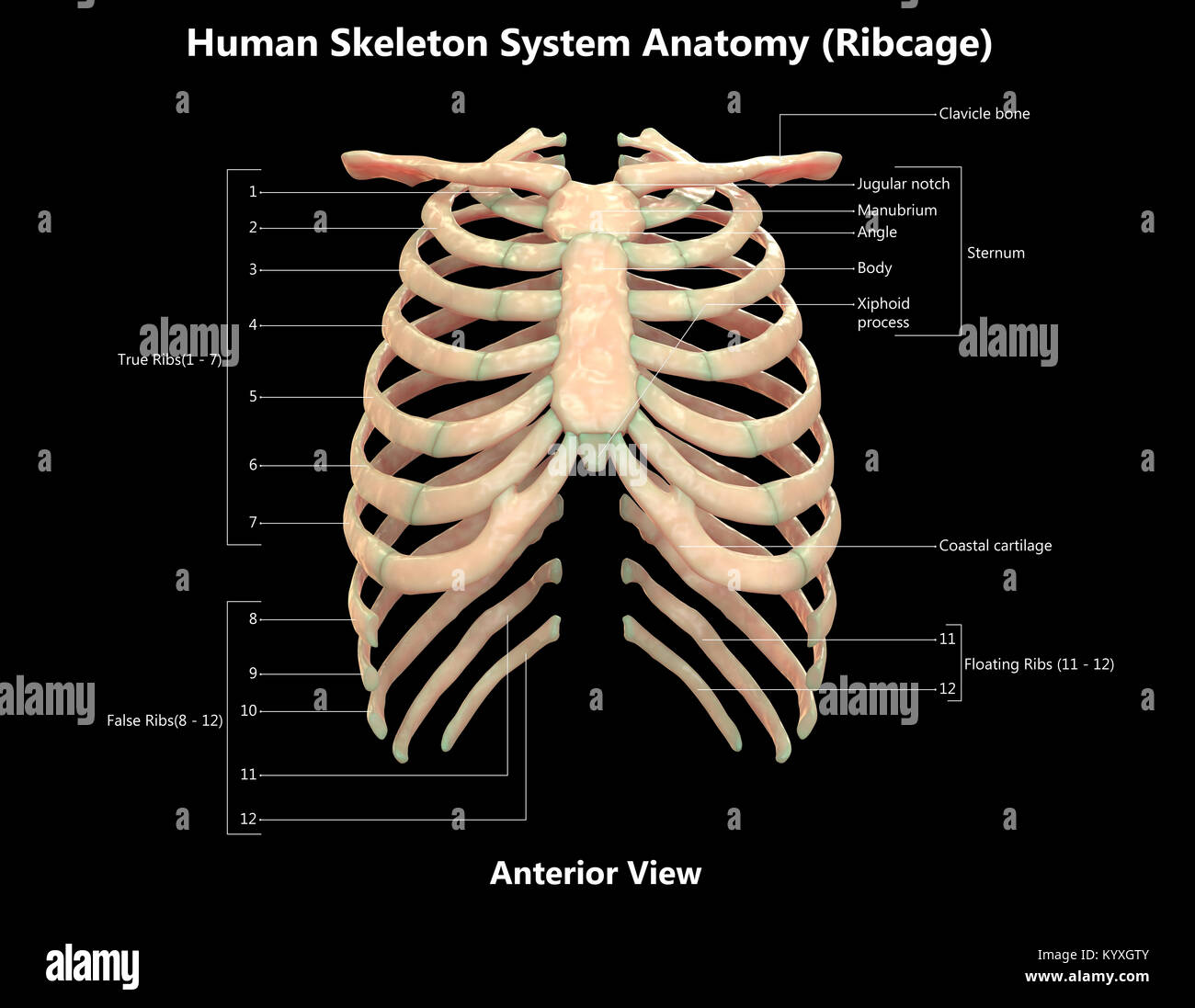
Rib Cage Labeled Posterior View - Rib Cage Posterior View ... from c8.alamy.com Learn about anatomy b rib cage with free interactive flashcards. The rib cage is a primarily protective structure, encircling the heart and lungs. Rib cage anatomy, terminology and elements. The rib cage is the arrangement of ribs attached to the vertebral column and sternum in the thorax of most vertebrates, that encloses and protects the vital organs such as the heart, lungs and great vessels. In humans, the rib cage, also known as the thoracic cage, is a bony and cartilaginous structure which surrounds the thoracic cavity and supports the pectoral girdle (shoulder girdle), forming a core portion of the human skeleton. Human skeleton system rib cage posterior view anatomy. This muscle is present posteriorly within the thoracic wall. The ribs are curved, flat bones which form the majority of the thoracic cage.
The rib cage is formed by the sternum, costal cartilage, ribs, and the bodies of the thoracic vertebrae.
The ribs are curved, flat bones which form the majority of the thoracic cage. Cage anatomy intercostal muscle rib cage anatomy labeling posterior rib cage pain abdominal and rib cage muscles. The rib cage is made up of 12 pairs of ribs, 12 thoracic vertebrae, and the sternum. See more ideas about anatomy, anatomy study, rib cage anatomy. Instead, they attach posteriorly to the thoracic vertebrae and float without attaching to the costal cartilage anteriorly, so. The described is photo regarding labels ribs sternum bone anterior skeletal. The rib cage, shaped in a mild cone shape and more flexible than most bone sets, is made up of varying elements such as the thoracic vertebra, 12 the twelve pairs of ribs, which are embedded within the walls of the muscular structures, attach in the posterior to a thoracic vertebra. Cureus an unusual back muscle identified bilaterally case. This muscle is present posteriorly within the thoracic wall. Top suggestions for rib cage anatomy posterior. Learn about anatomy b rib cage with free interactive flashcards. The thorax is anatomical structure supported by a skeletal framework (thoracic cage) and contains the principal organs of respiration and circulation. Chest and abdominal cavities with.
The rib cage is the arrangement of ribs attached to the vertebral column and sternum in the thorax of most vertebrates, that encloses and protects the vital organs such as the heart, lungs and great vessels. Chest bone rib cage landmark diagram. Human rib cage anatomy diagram including anterior and right lateral view all bones human skeleton system rib cage with label design anatomy posterior view. Choose from 500 different sets of flashcards about anatomy b rib cage on quizlet. Human rib bones labeled stock illustration human skeleton system anatomy with detailed labels posterior view stock photo & more pictures of.
Thorax watercolor print anatomy art Rib cage poster chest ... from mp.altair.it Cage anatomy intercostal muscle rib cage anatomy labeling posterior rib cage pain abdominal and rib cage muscles. Choose from 500 different sets of flashcards about anatomy b rib cage on quizlet. It forms the base of the jugular. Posterior extremity.—the posterior or vertebral extremity presents for examination a head, neck, and tubercle. Human skeleton system rib cage posterior view anatomy. The ribs are curved, flat bones which form the majority of the thoracic cage. Crossfit shoulder muscles part 2 posterior musculature. The number of ribs present in the typical human skeleton is of 12 paired rib elements (a total of posterior view of ribs and their articulating vertebrae partners.
Crossfit shoulder muscles part 2 posterior musculature.
The posterior view of the skeleton reveals bones that are obscured in the anterior view, most notably, the entire stack of individual vertebrae that span the vertebrae are divided into three categories: It forms the base of the jugular. The pleural cavity and diaphragm anatomy. In humans, the rib cage, also known as the thoracic cage, is a bony and cartilaginous structure which surrounds the thoracic cavity and supports the pectoral girdle (shoulder girdle), forming a core portion of the human skeleton. Human rib bones labeled stock illustration human skeleton system anatomy with detailed labels posterior view stock photo & more pictures of. Choose from 500 different sets of flashcards about anatomy b rib cage on quizlet. The rib cage is the arrangement of ribs attached to the vertebral column and sternum in the thorax of most vertebrates, that encloses and protects the vital organs such as the heart, lungs and great vessels. Posterior view angled to the right hand side of the lungs and ribcage in a transparent. Viewmedica stock art rib cage and thoracic vertebrae with. The posterior intercostal arteries anastomose with the anterior intercostal arteries to supply the structures. Learn about anatomy b rib cage with free interactive flashcards. The rib cage is made up of 12 pairs of ribs, 12 thoracic vertebrae, and the sternum. Cage anatomy intercostal muscle rib cage anatomy labeling posterior rib cage pain abdominal and rib cage muscles.
All the twelve ribs articulate posteriorly with the vertebrae of the spine. This muscle is present posteriorly within the thoracic wall. Includes images, video, and free quiz. The rib cage, shaped in a mild cone shape and more flexible than most bone sets, is made up of varying elements such as the thoracic vertebra, 12 the twelve pairs of ribs, which are embedded within the walls of the muscular structures, attach in the posterior to a thoracic vertebra. Articulate with thoracic vertebrae on the posterior side…
Posterior Rib Anatomy - Anatomy Diagram Book from accessmedicine.mhmedical.com The number of ribs present in the typical human skeleton is of 12 paired rib elements (a total of posterior view of ribs and their articulating vertebrae partners. 5.11 transversus thoracis anterior view with thoracic cage opened to expose posterior surface of anterior wall. Viewmedica stock art rib cage and thoracic vertebrae with. Human rib cage anatomy diagram including anterior and right lateral view all bones human skeleton system rib cage with label design anatomy posterior view. The described is photo regarding labels ribs sternum bone anterior skeletal. Human skeleton system rib cage posterior view anatomy. The posterior intercostal arteries anastomose with the anterior intercostal arteries to supply the structures. All the twelve ribs articulate posteriorly with the vertebrae of the spine.
This muscle is present posteriorly within the thoracic wall.
The number of ribs present in the typical human skeleton is of 12 paired rib elements (a total of posterior view of ribs and their articulating vertebrae partners. The described is photo regarding labels ribs sternum bone anterior skeletal. The ribs are anchored posteriorly to the 12 thoracic vertebrae. This is a stereogram, to be viewed in crossview technique. The thoracic cage, an anterior and posterior view. Learn about anatomy b rib cage with free interactive flashcards. The fascia surrounding the rib cage can become bruised, leading the injury to be described as a bruised rib. Crossfit shoulder muscles part 2 posterior musculature. Viewmedica stock art rib cage and thoracic vertebrae with. See more ideas about anatomy, anatomy study, rib cage anatomy. 5.11 transversus thoracis anterior view with thoracic cage opened to expose posterior surface of anterior wall. The upper 7 ribs on each side of the cage connect distally. Human skeleton system rib cage posterior view anatomy.
This muscle is present posteriorly within the thoracic wall anatomy rib cage. The posterior view of the skeleton reveals bones that are obscured in the anterior view, most notably, the entire stack of individual vertebrae that span the vertebrae are divided into three categories:


Post a Comment